غدة تحت اللسان
| غدة تحت اللسان | |
|---|---|
 الغدد اللعابية في الجانب الأيمن | |
 | |
| Details | |
| جزء من | الغدد اللعابية |
| الجهاز | Digestive system |
| الموقع | الفم |
| الشريان | Sublingual artery (branch of lingual artery of external carotid artery) |
| العصب | Submandibular ganglion |
| اللمف | Submandibular lymph nodes |
| المُعرفات | |
| اللاتينية | glandula sublingualis |
| المصطلحات التشريحية | |
الغدة تحت اللسان إنگليزية: Sublingual gland إحدى الغدد اللعابية، تقع إلى الأمام من الغدة تحت الفك السفلي تحت اللسان وذلك إلى العمق من الغشاء المخاطي لأرضية الفم. وهي الأصغر والأكثر تشتتاً بين الغدد اللعابية الرئيسية الثلاثة في التجويف الفموي، والأخريين هما submandibular and parotid. The sublingual gland provides approximately 3-5% of the total salivary volume.[1]
البنية
The sublingual glands are located anterior and superior to the submandibular gland and inferior and lateral to the tongue, as well as inferior to the mucous membrane of the floor of the mouth. They are bound laterally by the bone of the mandible and inferolaterally by the mylohyoid muscle. The glands can be palpated posteriorly to each mandibular canine. Placing one index finger within the mouth and the fingertips of the opposite hand outside it, the compressed gland is manually palpated between the inner and outer fingers.[مطلوب توضيح][2]
The sublingual gland is constituted by 1 major duct and approximately 20 small excretory ducts, with the latter often being referred to as ducts of Rivinus.[3] The largest of all, the sublingual duct (of Bartholin) joins the submandibular duct to drain through the sublingual caruncle. The sublingual caruncle is a small papilla near the midline of the floor of the mouth on each side of the lingual frenum.[2] Most of the remaining small sublingual ducts (of Rivinus) open separate into the mouth on an elevated crest of mucous membrane, the plica sublingualis (aka sublingual fold), formed by the gland and located on either side of the frenulum linguae.

التشريح الدقيق
The sublingual gland consists mostly of mucous acini having serous demilunes and is therefore categorized as a mixed mucous gland with mostly a mucous product. Striated and intercalated ducts are also present.[4]
الإمداد بالدم
The gland receives its blood supply from the sublingual and submental arteries.[3] Lymph from the sublingual salivary gland drains into the submandibular lymph nodes.[2]
الإمداد العصبي
The chorda tympani nerve (from the facial nerve via the submandibular ganglion) is secretomotor and provides parasympathetic supply to the sublingual glands. The path of the nerve is as follows: junction between pons and medulla, through internal acoustic meatus and facial canal to chorda tympani, through middle ear cavity, out petrotympanic fissure to join the lingual nerve, travels with lingual nerve to synapse at the submandibular ganglion, then postganglionic fibers travels to the sublingual gland.
النمو
The sublingual salivary glands appear in the eighth week of prenatal development, two weeks later than the other two major salivary glands. They develop from epithelial buds in the sulcus surrounding the sublingual folds on the floor of the mouth, lateral to the developing submandibular gland. These buds branch and form into cords that canalize to form the sublingual ducts associated with the gland. The rounded terminal ends of the cords form acini. [5]
الأهمية الإكلينيكية
Ranulas are the most common pathologic lesion associated with the sublingual glands.[6]
صور إضافية
-
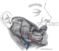 Imaging showing the sublingual glands and surrounding structures.
 -
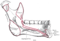 Mandible. Inner surface. Side view.
 -
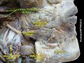 Sublingual gland
 -
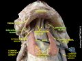 Sublingual gland
 -
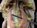 Sublingual gland


المراجع
- ^ Fehrenbach, MJ and Popowics, T. (2026). Illustrated Dental Embryology, Histology, and Anatomy, 6th edition, Elsevier, page 153.
- ^ أ ب ت Illustrated Anatomy of the Head and Neck, Fehrenbach and Herring, Elsevier, 2012, p. 156
- ^ أ ب Ten Cate's Oral Histology, Nanci, Elsevier, 2013, page 255
- ^ Fehrenbach, MJ and Popowics, T. (2026). Illustrated Dental Embryology, Histology, and Anatomy, 6th edition, Elsevier, page 153.
- ^ Fehrenbach, MJ and Popowics, T. (2026). Illustrated Dental Embryology, Histology, and Anatomy, 6th edition, Elsevier, pages 154-55.
- ^ Hupp JR, Ellis E, Tucker MR (2008). Contemporary oral and maxillofacial surgery (5th ed.). St. Louis, Mo.: Mosby Elsevier. pp. 410–411. ISBN 9780323049030.
وصلات خارجية
- SUNY Labs 34:st-0701 - "Oral Cavity: Glands"
- Norman/Georgetown cranialnerves (VII)
- "Anatomy diagram: 25420.000-1". Roche Lexicon - illustrated navigator. Elsevier. Archived from the original on 2015-02-26.
- Salivary gland infections from Medline Plus
- Salivary gland cancer from American Cancer Society
