وريد أجوف سفلي
| الوريد: الوريد الأجوف السفلي Inferior vena cava | |
|---|---|
 | |
| Anterior (frontal) view of the opened heart. White arrows indicate normal blood flow. | |
 | |
| Superior vena cava, inferior vena cava, azygos vein and their tributaries. | |
| اللاتينية | vena cava inferior |
| گراي | subject #173 677 |
| المصدر | common iliac vein lumbar veins testicular vein renal vein suprarenal vein hepatic vein |
| يصرّف إلى | heart |
| الشريان | abdominal aorta |
| MeSH | Vena+Cava,+Inferior |
الوريد الأجوف السفلي inferior vena cava وعاء دموي يقوم بحمل الدم من الأحشاء والطرفين السفليين إلى القلب .
The caval opening is at T8. The specific levels of the tributaries are as follows:
| الوريد | المستوى |
| inferior phrenic vein | T8 |
| hepatic veins | T8 |
| suprarenal vein | L1 |
| renal veins | L1 |
| gonadal vein | L2 |
| lumbar veins | L1-L5 |
| common iliac veins | L5 |
صور إضافية
-
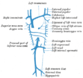 Diagram showing completion of development of the parietal veins.
 -
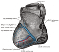 Base and diaphragmatic surface of heart.
 -
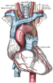 The arch of the aorta, and its branches.
 -
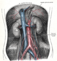 The abdominal aorta and its branches.
 -
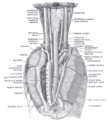 The position and relation of the esophagus in the cervical region and in the posterior mediastinum. Seen from behind.
 -
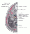 Transverse section of human embryo eight and a half to nine weeks old.
 -
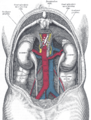 Human kidneys viewed from behind with spine removed
 -
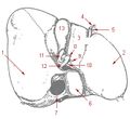 Liver and gallbladder
 -
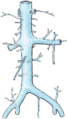 Inferior vena cava


انظر أيضاً
الهامش
وصلات خارجية
- synd/1462 at Who Named It? - "Eustachian valve"
- SUNY Labs 40:13-0101 - "Posterior Abdominal Wall: Tributaries to the Inferior Vena Cava"
- SUNY Anatomy Image 8827
- Cross section at UV pembody/body12a

 | هذه بذرة مقالة عن علم التشريح تحتاج للنمو والتحسين، ساهم في إثرائها بالمشاركة في تحريرها. |